Santiago Cajal Doctor of Medicine, Ph.D.
Santiago Cajal Doctor of Medicine, Ph.D.
The Nobel Prize in Physiology or Medicine 1906
Co-nobelist: Camillo Golgi
Name: Santiago Ramón y Cajal
Birth: 1 May 1852, Petilla de Aragón, Spain
Death: 17 October 1934, Madrid, Spain
Affiliation: Madrid University, Madrid, Spain
Award: “in recognition of their work on the structure of the nervous system.”
Portion of Cash: 1/2
Education: Doctor of Medicine, Zaragoza, Madrid, 1873. Ph.D., University of Zaragoza, 1877.
“Oh comforting solitude, how favorable thou art to original thought!”
“Outstanding work results from immense zeal applied to great idea.”
Nobel Prize Cash & Philanthropy
Images
Videos
Images
Historic photograph of a young Santiago Ramon y Cahal in a research laboratory. © Dr Mark Hill 2016, UNSW Embryology ISBN: 978 0 7334 2609 4 – UNSW CRICOS Provider Code No. 00098G
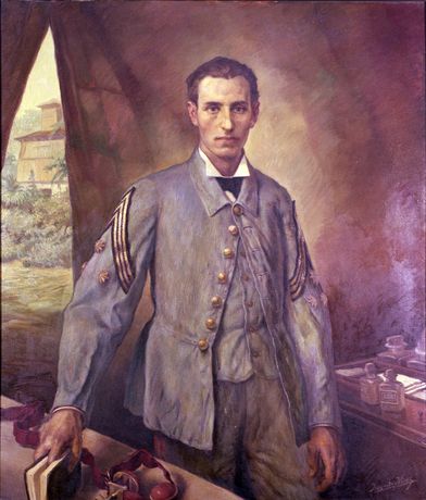
The Captain Doctor Santiago Ramón y Cajal Portrait, in 1874. Ten Years’ War in Cuba. Oil on canvas. Army museum, Spain.
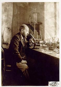
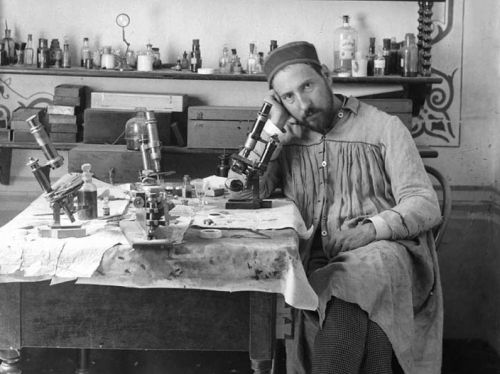
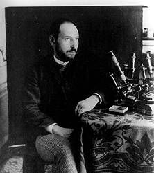
Self-portrait looking through a microscope.
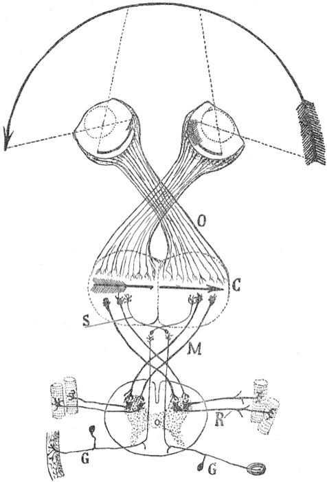
Schema of the visual map theory (1898). O=Optic chiasm; C=Visual (and motor) cortex; M, S=Decussating pathways; R, G: Sensory nerves, motor ganglia.
A schematic illustration of a section of the spinal cord depicting the nerve roots. Illustration by Cajal from the book Recuerdos de mi vida.
U.S National Library of Medicine, History of Medicine Division
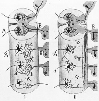
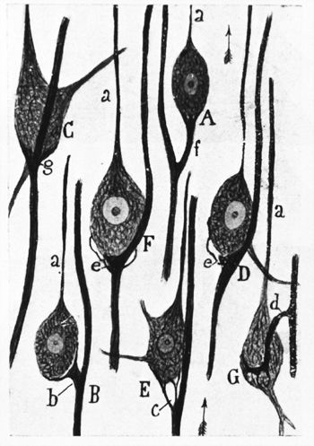
Neuroglial cells of a cat. Illustration by Cajal from the book Recuerdos de mi vida. U.S National Library of Medicine, History of Medicine Division
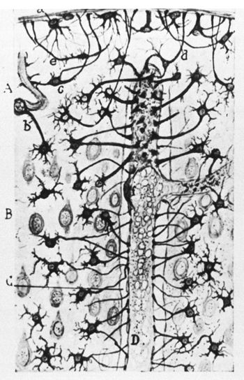
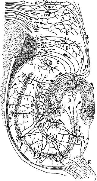
Detail of neural connections. Illustration by Cajal from the book Recuerdos de mi vida. U.S National Library of Medicine, History of Medicine Division.
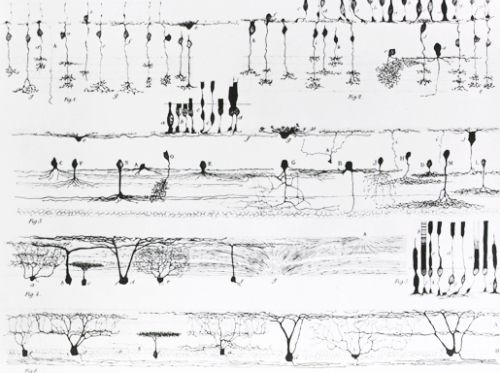
Various types of nerve structures. Illustration by Cajal from the book Die Retina der Wirbelthiere, Tafel II. U.S National Library of Medicine, History of Medicine Division.
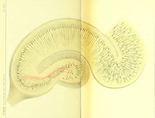
Drawing of the neural circuitry of the rodent hippocampus. By Santiago Ramón y Cajal. Histologie du Systeme Nerveux de l’Homme et des Vertebretes, Vols. 1 and 2. A. Maloine. Paris. 1911, Wiki.
Golgi’s drawing of the hippocampus, based on tissues he had stained. From Golgi’s 1886 publication “Sulla fina anatomia degli organi centrali del sistema nervoso.”
Drawing of a section through the optic tectum of a sparrow, from “Estructura de los centros nerviosos de las aves”, Madrid, 1905, Wiki.
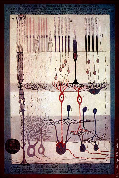
“Structure of the Mammalian Retina” Madrid, 1900, Wiki.
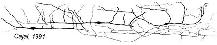
Drawing of Cajal-Retzius cells, 1891
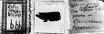
Microscope slides with Cajal’s histological preparations; the letter ‘b’ (bueno, good) indicates the quality of the sections.
From book “Santiago Ramón y Cajal o la Pasión de España” by Agustín Albarracín, published by Editorial Labor, S.A. 1982.
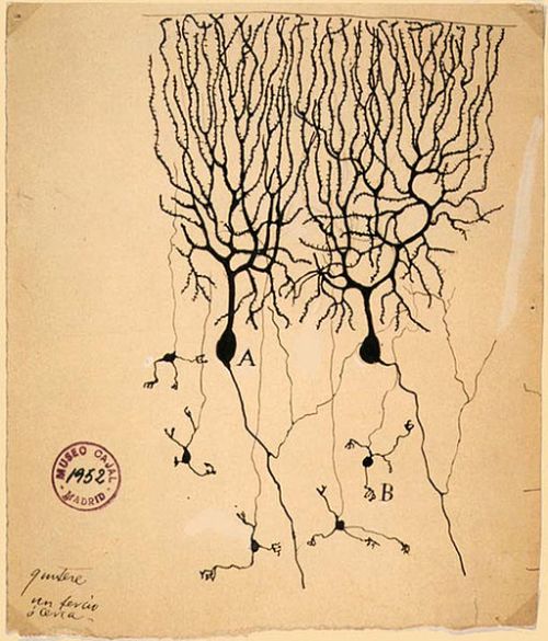
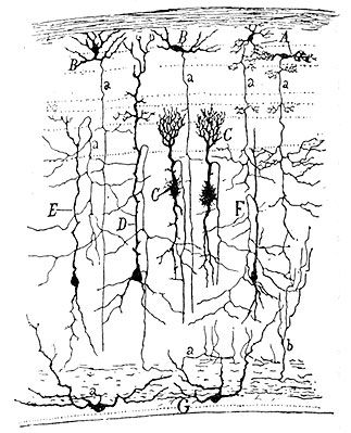
899 drawing of Purkinje cells (A) and granule cells (B) from pigeon cerebellum by Santiago Ramón y Cajal.image © Instituto Cajal, Madrid, Spain
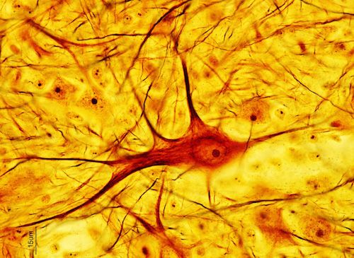
Neuron cell structures dyed with the “Golgi stain” method. Source NIH.
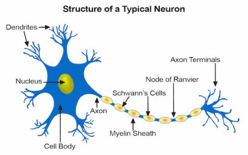
A typical neuron structure consisting of three parts: a cell body (soma), one or more dendrites, and an axon. Image NIH
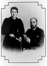
Portrait of Cajal with his wife in their first years in Madrid.
From book “Santiago Ramón y Cajal o la Pasión de España” by Agustín Albarracín, published by Editorial Labor, S.A. 1982.
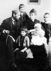
Self-portrait of Cajal with his children (from left to right: Fe, Jorge, Pula and Santiago) in Barcelona. From book “Santiago Ramón y Cajal o la Pasión de España” by Agustín Albarracín, published by Editorial Labor, S.A. 1982.
Self-portrait of Cajal in his laboratory in Valencia. From book “Santiago Ramón y Cajal o la Pasión de España” by Agustín Albarracín, published by Editorial Labor, S.A. 1982.
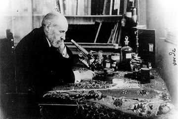
Self-portrait of Cajal at the microscope in 1920. From book “Santiago Ramón y Cajal o la Pasión de España” by Agustín Albarracín, published by Editorial Labor, S.A. 1982.
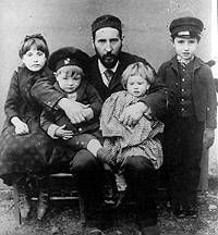
Santiago Ramon y Cajal with his family, 1890. © Nigel Hunt.
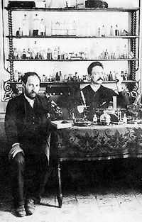
Santiago Ramon y Cajal (left) with an assistant in his laboratory in Valencia, 1884. © Nigel Hunt.
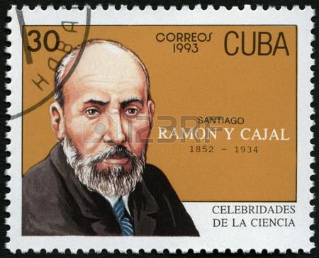
Cuban stamp, 1993: Santiago Ramon y Cajal, histologist. Source: 123rf website.
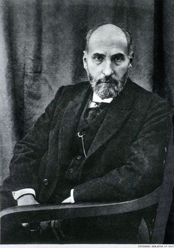
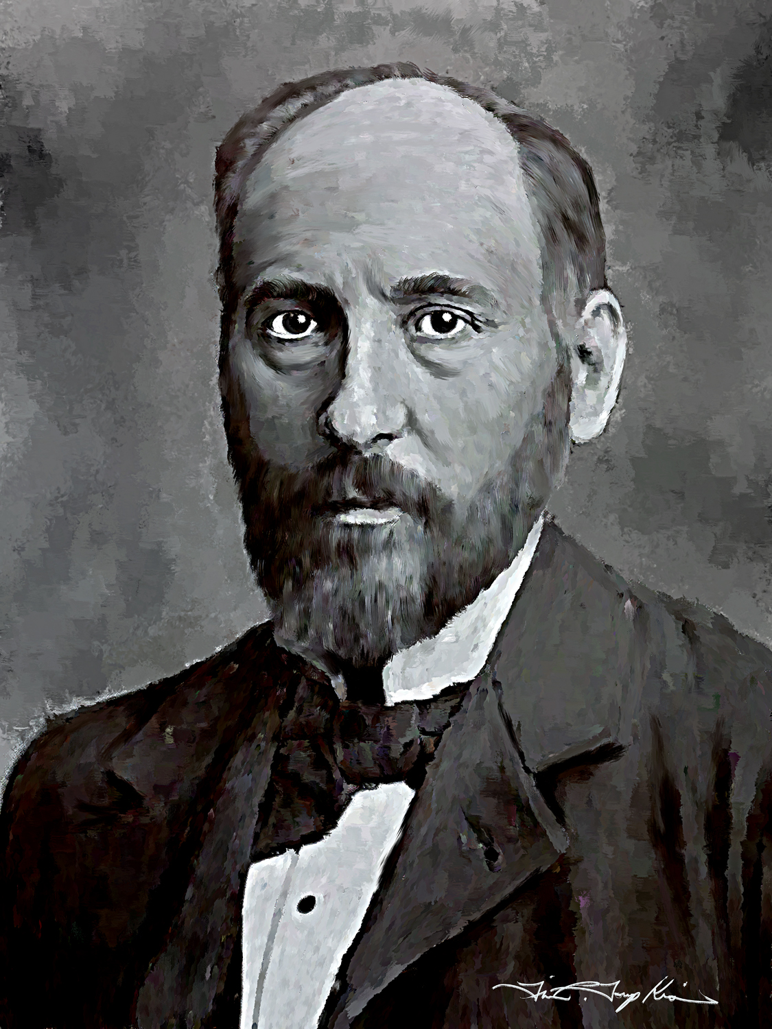
Portrait of Santiago Ramón y Cajal. U.S National Library of Medicine, History of Medicine Division.
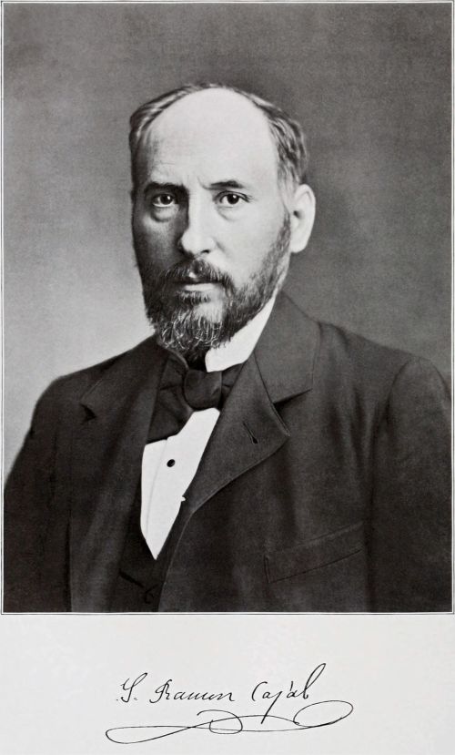
Santiago Ramón y Cajal. Source: Clark University, 1899. Restoration by Garrondo. Wiki.
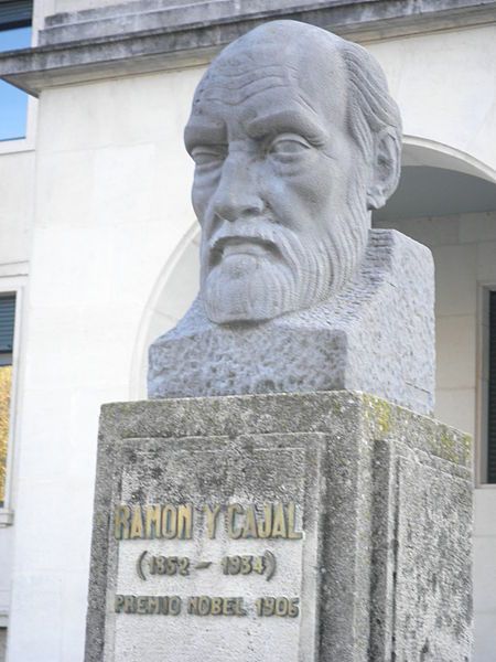
Photo HCPUNXKID, Wiki.